Diagnosing rheumatic diseases can be difficult because some symptoms and signs are common to many different diseases. A general practitioner or family doctor may be able to evaluate a patient or refer him or her to a rheumatologist (a doctor who specializes in treating arthritis and other rheumatic diseases).
Common Signs and Symptoms of Arthritis
- swelling in one or more joints
- stiffness around the joints that lasts for at least 1 hour in the early morning
- constant or recurring pain or tenderness in a joint
- difficulty using or moving a joint normally
- warmth and redness in a joint
The doctor will review the patient’s medical history, conduct a physical examination, and obtain laboratory tests and x rays or other imaging tests. The doctor may need to see the patient more than once and possibly a number of times to make an accurate diagnosis.
Medical History
It is vital for people with joint pain to give the doctor a complete medical history. Answers to the following questions will help the doctor make an accurate diagnosis:
- Is the pain in one or more joints?
- When does the pain occur?
- How long does the pain last?
- When did you first notice the pain?
- What were you doing when you first noticed the pain?
- Does activity make the pain better or worse?
- Have you had any illnesses or accidents that may account for the pain?
- Are you experiencing any other symptoms besides pain?
- Is there a family history of arthritis or other rheumatic disease?
- What medicine(s) are you taking?
- Have you had any recent infections?
Because rheumatic diseases are so diverse and sometimes involve several parts of the body, the doctor may ask many other questions.
It may be helpful for people to keep a daily journal that describes the pain. Patients should write down what the affected joint looks like, how it feels, how long the pain lasts, and what they were doing when the pain started.
Physical Examination and Laboratory Tests
The doctor will examine the patient’s joints for redness, warmth, damage, ease of movement, and tenderness. Because some forms of arthritis, such as lupus, may affect internal organs, a complete physical examination that includes the heart, lungs, abdomen, nervous system, eyes,
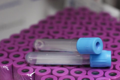
ears, mouth, and throat may be necessary. The doctor may order some laboratory tests to help confirm a diagnosis. Samples of blood, urine, or synovial fluid (lubricating fluid found in the joint) may be needed for the tests. Many of these same tests may be useful later for monitoring the disease or the effectiveness of treatments.
Common laboratory tests and procedures include the following:
Antinuclear antibody (ANA). This test checks blood levels of antibodies that are often present in people who have connective tissue diseases or other autoimmune disorders, such as lupus. Because the antibodies react with material in the cell’s nucleus (control center), they are referred to as antinuclear antibodies. There are also tests for individual types of ANAs that may be more specific to people with certain autoimmune disorders. ANAs are also sometimes found in people who do not have an autoimmune disorder. (In such cases, the result is referred to as a “false positive.”) Therefore, having ANAs in the blood does not necessarily mean that a person has a disease.
CCP (or anti-CCP). This test checks blood levels of antibodies to citrulline, a protein that can be detected in up to 70 percent of people in the early stages of rheumatoid arthritis. Because the presence of anti-CCPs is associated with more aggressive disease, the test can also be useful in helping doctors plan treatment.
C-reactive protein test. This nonspecific test is used to detect generalized inflammation. Levels of the protein are often increased in patients with active disease such as rheumatoid arthritis or any other disease that causes inflammation.
Complement. This test measures the level of complement, a group of proteins in the blood. Complement helps destroy germs and other foreign substances that enter the body. A low blood level of complement is common in people who have active lupus.
Complete blood count (CBC). This test determines the number of white blood cells, red blood cells, and platelets present in a sample of blood. Some rheumatic conditions or drugs used to treat arthritis are associated with a low white blood count (leukopenia), low red blood count (anemia), or low platelet count (thrombocytopenia).
Creatinine. This blood test measures the level of creatinine, a breakdown product of creatine, which is an important component of muscle. Creatinine is excreted from the body entirely by the kidneys, and the level remains constant and normal when kidney function is normal. This test is commonly used to diagnose and monitor kidney disease in patients who have a rheumatic condition such as lupus.
Erythrocyte sedimentation rate (sed rate or ESR). This blood test is used to detect inflammation in the body. Higher sed rates, indicating the presence of inflammation, are typical of many forms of arthritis, such as rheumatoid arthritis and ankylosing spondylitis. Higher sed rates are also typical of many of the immunologic connective tissue diseases, such as lupus and scleroderma.
Hematocrit (PCV or packed cell volume). This test and the test for hemoglobin (a substance in the red blood cells that carries oxygen throughout the body) measure the number of red blood cells present in a sample of blood. A decrease in the number of red blood cells (anemia) is common in people who have inflammatory arthritis or another rheumatic disease.
Rheumatoid factor. This test detects the presence of rheumatoid factor, an antibody found in the blood of most (but not all) people who have rheumatoid arthritis. In rheumatoid arthritis, it is associated with more aggressive disease. Rheumatoid factor may be found in many diseases besides rheumatoid arthritis and sometimes in people without health problems. (In the latter case, the result is referred to as a “false positive.”)
Synovial fluid examination. Synovial fluid may be examined for white blood cells (found in patients with rheumatoid arthritis and infections), bacteria or viruses (found in patients with infectious arthritis), or crystals in the joint (found in patients with gout or other types of crystal-induced arthritis). To obtain a specimen, the doctor injects a local anesthetic, then inserts a needle into the joint to withdraw the synovial fluid into a syringe. The procedure is called arthrocentesis or joint aspiration.
Urinalysis. In this test, a urine sample is studied for protein, red blood cells, white blood cells, and bacteria. These abnormalities may indicate kidney disease, which may be seen in lupus as well as several rheumatic conditions. Some medications used to treat arthritis also can cause abnormal findings on urinalysis.
X Rays and Other Imaging Procedures
To see what the joint looks like inside, the doctor may order x rays or other imaging procedures. X rays provide an image of the bones, but they do not show cartilage, muscles, and ligaments. Other noninvasive imaging methods such as computed tomography (CT or CAT scan), magnetic resonance imaging (MRI), and arthrography show the whole joint. The doctor also may look for damage to a joint by using an arthroscope, a small, flexible tube that is inserted through a small incision at the joint. The arthroscope transmits the image from inside the joint to a video screen.
